

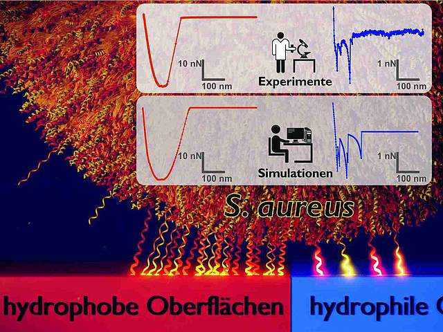
Bacterial adhesion to surfaces is a crucial step in initial biofilm formation. In a combined experimental and computational approach, the groups of Karin Jacobs and Markus Bischoff (B2) and the group of Ludger Santen (A8) studied the adhesion of the pathogenic bacterium Staphylococcus aureus to hydrophilic and hydrophobic surfaces. They used atomic force microscopy-based single-cell force spectroscopy and Monte Carlo simulations to investigate the similarities and differences of adhesion to hydrophilic and hydrophobic surfaces. Their results reveal that binding to both types of surfaces is mediated by thermally fluctuating cell wall macromolecules that behave differently on each type of substrate: on hydrophobic surfaces, many macromolecules are involved in adhesion, yet only weakly tethered, leading to high variance between individual bacteria, but low variance between repetitions with the same bacterium. On hydrophilic surfaces, however, only few macromolecules tether strongly to the surface. Since during every repetition with the same bacterium different macromolecules bind, they observed a comparable variance between repetitions and different bacteria. These findings are expected to be of importance for the understanding of the adhesion behaviour of many bacterial species as well as other microorganisms and even nanoparticles with soft, macromolecular coatings, used e.g. for biological diagnostics.
 |
 |
 |
| Model of the adhesion mechanism by which the bacterium Staphylococcus aureus binds to hydrophobic (‘low-energy’) surfaces (left) compared with hydrophilic (‘high-energy’) surfaces (right). On the left, a large number of cell wall molecules (shown here as tiny compressible springs) are involved in binding the cell to the hydrophobic surface. On the hydrophilic surface shown on the right, far fewer molecules are involved. The results were obtained by a team of experimental and theoretical physicists at Saarland University who performed computational Monte Carlo simulations of force-distance data from atomic force microscopy experiments. |
© Universität des Saarlandes
Prof. Dr. Karin Jacobs
|
© Universität des Saarlandes
Prof. Dr. Ludger Santen
|
Link to the publication - click here
Press release of the Saarland University - in German / in English
Press release of the IDW - click here